Back to Profile Page
Tree of Life Media Contributed By David J. Patterson
<<
<
3
4
5
6
7
8
9
>
>>
|
ID
|
Thumbnail |
Media Data |
| 31238 |
|
|
Scientific Name
|
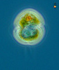
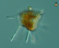
Karlodinium micrum
|
|
Comments
|
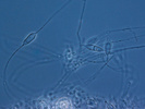
Karlodinium micrum has an equatorial flagellum lying in a groove (girdle or cingulum) near the centre of the cell and a second flagellum trailing behind the cell and arising in a longitudinal groove or sulcus. The large gray area posterior to the girdle is the nucleus. The orange element is probably a residue from ingested food (Rhodomonas).
|
|
Acknowledgements
|
This image is of material from Provasoli-Guillard National Center for Culture of Marine Phytoplankton
|
|
Source
|
Karlodinium micrum
|
|
Source Collection
|
Micro*scope
|
|
Image Use
|
 This media file is licensed under the Creative Commons Attribution-NonCommercial-ShareAlike License - Version 2.5. This media file is licensed under the Creative Commons Attribution-NonCommercial-ShareAlike License - Version 2.5.
|
|
Copyright
|
© Bob Andersen and D. J. Patterson
|
|
Attached to Group
|
Karlodinium (Haptophores): view page image collection
|
|
Title
|
gyrodinium_bsa.jpg
|
|
Image Type
|
Photograph
|
|
Image Content
|
Specimen(s)
|
|
Technical Information
|
Phase contrast microscopy.
|
|
ID
|
31238
|
|
| 31239 |
|
|
Scientific Name
|
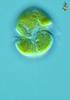
Karlodinium micrum
|
|
Comments
|
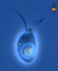
Karlodinium micrum has an equatorial flagellum lying in a groove (girdle or cingulum) near the centre of the cell and a second flagellum trailing behind the cell and arising in a longitudinal groove or sulcus. The grooves and plastids are emphasized in this image.
|
|
Acknowledgements
|
This image is of material from Provasoli-Guillard National Center for Culture of Marine Phytoplankton
|
|
Source
|
Karlodinium micrum
|
|
Source Collection
|
Micro*scope
|
|
Image Use
|
This media file is licensed under the Creative Commons Attribution-NonCommercial-ShareAlike License - Version 2.5.
|
|
Copyright
|
© Bob Andersen and D. J. Patterson
|
|
Attached to Group
|
Karlodinium (Haptophores): view page image collection
|
|
Title
|
gyrodinium2_bsa.jpg
|
|
Image Type
|
Photograph
|
|
Image Content
|
Specimen(s)
|
|
Technical Information
|
Differential interference microscopy
|
|
ID
|
31239
|
|
| 31240 |
|
|
Scientific Name
|
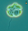
Karenia brevis
|
|
Comments
|
Karenia brevis, small gymnodinioid dinoflagellate, cell with equatorial groove or girdle containing one flagellum, a second flagellum trails behind the cell. With golden chloroplasts, and large globular nucleus in posterior part of the cell.
|
|
Acknowledgements
|
This image is of material from Provasoli-Guillard National Center for Culture of Marine Phytoplankton
|
|
Source
|
Karenia brevis
|
|
Source Collection
|
Micro*scope
|
|
Image Use
|
This media file is licensed under the Creative Commons Attribution-NonCommercial-ShareAlike License - Version 2.5.
|
|
Copyright
|
© Bob Andersen and D. J. Patterson
|
|
Attached to Group
|
Karenia (Haptophores): view page image collection
|
|
Title
|
karebiabrevis_bga.jpg
|
|
Image Type
|
Photograph
|
|
Image Content
|
Specimen(s)
|
|
Technical Information
|
Phase contrast microscopy
|
|
ID
|
31240
|
|
| 31241 |
|
|
Scientific Name
|
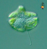
Karenia brevis
|
|
Comments
|
Karenia brevis, small gymnodinioid dinoflagellate, cell with equatorial groove or girdle containing one flagellum, a second flagellum trails behind the cell. With golden chloroplasts, and large globular nucleus in posterior part of the cell, in this image showing the condensed chromosomes.
|
|
Acknowledgements
|
This image is of material from Provasoli-Guillard National Center for Culture of Marine Phytoplankton
|
|
Source
|
Karenia brevis
|
|
Source Collection
|
Micro*scope
|
|
Image Use
|
This media file is licensed under the Creative Commons Attribution-NonCommercial-ShareAlike License - Version 2.5.
|
|
Copyright
|
© Bob Andersen and D. J. Patterson
|
|
Attached to Group
|
Karenia (Haptophores): view page image collection
|
|
Title
|
karebiabrevis2_bga.jpg
|
|
Image Type
|
Photograph
|
|
Image Content
|
Specimen(s)
|
|
Technical Information
|
Differential interference microscopy
|
|
ID
|
31241
|
|
| 31243 |
|
|
Scientific Name
|
Ceratocorys horrida
|
|
Comments
|
Body armoured with projecting spines, some with wings. The girdle is edged with two projecting and ribbed flanges.
|
|
Acknowledgements
|
This image is of material from Provasoli-Guillard National Center for Culture of Marine Phytoplankton
|
|
Source
|
Ceratocorys horrida
|
|
Source Collection
|
Micro*scope
|
|
Image Use
|
This media file is licensed under the Creative Commons Attribution-NonCommercial-ShareAlike License - Version 2.5.
|
|
Copyright
|
© Bob Andersen and D. J. Patterson
|
|
Attached to Group
|
Ceratocorys: view page image collection
|
|
Title
|
ceratocorys_bga.jpg
|
|
Image Type
|
Photograph
|
|
Image Content
|
Specimen(s)
|
|
Technical Information
|
Differential interference microscopy
|
|
ID
|
31243
|
|
| 31262 |
|
|
Scientific Name
|
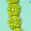
Gymnodinium catenatum
|
|
Comments
|
Gymnodinium catenatum is a chain forming motile species associated with the production of toxins.
|
|
Acknowledgements
|
This image is of material from Provasoli-Guillard National Center for Culture of Marine Phytoplankton
|
|
Body Part
|
This image of the ventral surface of several cells shows the equatorial groove or girdle and longitudinal groove or cingulum. Flagella can also be seen.
|
|
Source
|
Gymnodinium catenatum
|
|
Source Collection
|
Micro*scope
|
|
Image Use
|
This media file is licensed under the Creative Commons Attribution-NonCommercial-ShareAlike License - Version 2.5.
|
|
Copyright
|
© Bob Andersen and D. J. Patterson
|
|
Attached to Group
|
Gymnodiniaceae: view page image collection
|
|
Title
|
alexcatenella19373_bga.jpg
|
|
Image Type
|
Photograph
|
|
Image Content
|
Specimen(s)
|
|
Technical Information
|
Differential interference microscopy
|
|
ID
|
31262
|
|
| 31263 |
|
|
Scientific Name
|
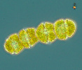
Gymnodinium catenatum
|
|
Comments
|
Gymnodinium catenatum is a chain forming motile species associated with the production of toxins.
|
|
Acknowledgements
|
This image is of material from Provasoli-Guillard National Center for Culture of Marine Phytoplankton
|
|
Body Part
|
Girdle (equatorial grooves) visible
|
|
Source
|
Gymnodinium catenatum
|
|
Source Collection
|
Micro*scope
|
|
Image Use
|
This media file is licensed under the Creative Commons Attribution-NonCommercial-ShareAlike License - Version 2.5.
|
|
Copyright
|
© Bob Andersen and D. J. Patterson
|
|
Attached to Group
|
Gymnodiniaceae: view page image collection
|
|
Title
|
alexcatenella19372_bga.jpg
|
|
Image Type
|
Photograph
|
|
Image Content
|
Specimen(s)
|
|
Technical Information
|
Phase contrast microscopy
|
|
ID
|
31263
|
|
| 31264 |
|
|
Scientific Name
|
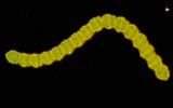
Gymnodinium catenatum
|
|
Comments
|
Gymnodinium catenatum is a chain forming motile species associated with the production of toxins. This chain contains 16 cells, but the chains may be of differing lengths, and other genera may form similar chains. Cells coloured because of presence of chloroplasts.
|
|
Acknowledgements
|
This image is of material from Provasoli-Guillard National Center for Culture of Marine Phytoplankton
|
|
Body Part
|
Girdle (equatorial grooves) visible
|
|
Source
|
Gymnodinium catenatum
|
|
Source Collection
|
Micro*scope
|
|
Image Use
|
This media file is licensed under the Creative Commons Attribution-NonCommercial-ShareAlike License - Version 2.5.
|
|
Copyright
|
© Bob Andersen and D. J. Patterson
|
|
Attached to Group
|
Gymnodiniaceae: view page image collection
|
|
Title
|
alexcatenella1937_bga.jpg
|
|
Image Type
|
Photograph
|
|
Image Content
|
Specimen(s)
|
|
Technical Information
|
Dark ground illumination
|
|
ID
|
31264
|
|
| 31334 |
|
|
Scientific Name
|
Cladochytrium sp.
|
|
Location
|
material from freshwater sites in the vicinity of the Universit y of Georgia in Athens, Georgia, USA and from collections of organisms maintained at the University
|
|
Comments
|
Cladochytrium is a chytrid, in which the cytoplasm develops into a system of fine branching rhizoids which are used to pick up food. This image shows the distinctive two part expansions of the rhizoidal system.
|
|
Source
|
Cladochytrium
|
|
Source Collection
|
Micro*scope
|
|
Image Use
|
This media file is licensed under the Creative Commons Attribution-NonCommercial-ShareAlike License - Version 2.5.
|
|
Copyright
|
© 2001 D. J. Patterson and Mark Farmer
|
|
Creation Date
|
April, 2001
|
|
Attached to Group
|
Chytridiales: view page image collection
|
|
Title
|
cladochytrium_uga.jpg
|
|
Image Type
|
Photograph
|
|
Image Content
|
Specimen(s)
|
|
ID
|
31334
|
|
| 31746 |
|
|
Please note: Most images and other media displayed on the Tree of Life web site are protected by copyright, and the ToL cannot act as an agent for their distribution. If you would like to use any of these materials for your own projects, you need to ask the copyright owner(s) for permission. For additional information, please refer to the
ToL Copyright Policies.

